


ņä£ļĪĀ
ļæÉĻ▓ĮļČĆņŚÉņä£ Ļ░Ćņן ĒØöĒĢ£ ņĢģņä▒ ņóģņ¢æņØĆ ĒÄĖĒÅē ņäĖĒżņĢöņØ┤ņ¦Ćļ¦ī, ņĢģņä▒ ļ”╝ĒöäņóģļÅä ņóģņóģ ļæÉĻ▓ĮļČĆ ņśüņŚŁņŚÉņä£ ļ░£ņāØĒĢ£ļŗż.1) ņĢģņä▒ ļ”╝ĒöäņóģņØĆ ĒśĖņ¦ĆĒé© ļ”╝ĒöäņóģĻ│╝ ļ╣äĒśĖņ¦ĆĒé© ļ”╝Ēöäņóģņ£╝ļĪ£ ļéśļłäņ¢┤ ņ¦ĆļŖöļŹ░, ņØ┤ ņżæ ļ╣äĒśĖņ¦ĆĒé© ļ”╝ĒöäņóģņØ┤ ļŹö ĒØöĒĢśļ®░, ļæÉĻ▓ĮļČĆņŚÉņä£ ļ░£ņāØĒĢśļŖö ņĢģņä▒ ņóģņ¢æņØś ļīĆļץ 5% ņĀĢļÅäļź╝ ņ░©ņ¦ĆĒĢ£ļŗż.2) ĒśĖņ¦ĆĒé© ļ”╝ĒöäņóģņØĆ ņŻ╝ļĪ£ Ļ▓ĮļČĆņÖĆ ņóģĻ▓®ļÅÖņØś ļ”╝ĒöäņĀłņŚÉņä£ ļ░£ņāØĒĢśļ®░, ļ”╝ĒöäņĀł ņÖĖ ļČĆņ£äņŚÉņä£ļŖö 5% ņĀĢļÅäņŚÉņä£ļ¦ī ļ░£ņāØĒĢśļŖöļŹ░ ļ░śĒĢ┤, ļ╣äĒśĖņ¦ĆĒé© ļ”╝ĒöäņóģņØĆ ļ”╝ĒöäņĀł ņÖĖ ļČĆņ£äņŚÉņä£ļÅä 30% ņĀĢļÅäĻ░Ć ļ░£ņāØĒĢśĻ│Ā, ļæÉĻ▓ĮļČĆņŚÉņä£ļŖö ņ╣©ņāś, ļČĆļ╣äļÅÖ, ļ░£ļŗżņØ┤ņ¢┤ ĒÄĖļÅä Ļ│Āļ”¼ ļō▒ņŚÉņä£ ņŻ╝ļĪ£ ļ░£ņāØĒĢśĻ│Ā ĻĘĖ ņóģļźśļŖö ļ»Ėļ¦īņä▒Ļ▒░ļīĆ BņäĖĒżļ”╝ĒöäņóģņØ┤ ņĀ£ņØ╝ ĒØöĒĢśļŗż.3,4) ļ░śļ®┤ ĒĢśņØĖļæÉ ļ░Å ĒøäļæÉņŚÉņä£ļŖö ņāüļīĆņĀüņ£╝ļĪ£ ļ”╝ĒöäņóģņØś ļ░£ņāØņØ┤ ļé«ļŗż. ĻĄŁļé┤ņŚÉņä£ļŖö ĒøäļæÉņŚÉņä£ ļ░£ņāØĒĢ£ ņĢģņä▒ļ”╝ĒöäņóģņØ┤ ļ│┤Ļ│ĀļÉ£ ņĀüņØĆ ņ׳ņ£╝ļéś,5) ĒĢśņØĖļæÉņŚÉņä£ ļ░£ņāØĒĢ£ ņĢģņä▒ļ”╝ĒöäņóģņØĆ ņĢäņ¦ü ļ│┤Ļ│ĀļÉ£ ļ░öĻ░Ć ņŚåļŗż. ĒŖ╣Ē׳ ĒĢśņØĖļæÉņŚÉņä£ļ¦ī ņøÉļ░£ņä▒ņ£╝ļĪ£ ļ░£ņāØĒĢ£ ņĀÉļ¦ē ņŚ░Ļ┤Ćņä▒ ļ”╝ĒöäņĪ░ņ¦ü ļ”╝ĒöäņóģņØĆ ņäĖĻ│äņĀüņ£╝ļĪ£ļÅä ļ¦żņÜ░ ļō£ļ¼╝ļŗż.6,7) ņĀĆņ×ÉļōżņØĆ 68ņäĖ ņŚ¼ņä▒ņŚÉņä£ ĒĢśņØĖļæÉņŚÉņä£ ļ░£ņāØĒĢ£ ņĀÉļ¦ē ņŚ░Ļ┤Ćņä▒ ļ”╝ĒöäņĪ░ņ¦ü ļ”╝Ēöäņóģ ņ”ØļĪĆļź╝ Ļ▓ĮĒŚśĒĢśņŚ¼ ļ│┤Ļ│ĀĒĢśļŖö ļ░öņØ┤ļŗż.
ņ”ØļĪĆ
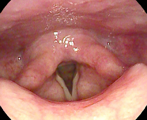
68ņäĖ ņŚ¼ņ×É ĒÖśņ×ÉĻ░Ć ĒĢ£ ļŗ¼ ņĀäļČĆĒä░ ļ░£ņāØĒĢ£ ļ¬® ņØ┤ļ¼╝Ļ░É ņŻ╝ņåīļĪ£ ņÖĖļל ļé┤ņøÉĒĢśņśĆļŗż. ĒØĪņŚ░ ļ░Å ņØīņŻ╝ļĀźņØĆ ņŚåņŚłņ£╝ļ®░, Ļ│╝Ļ▒░ļĀźņāü ņóīņĖĪ ņ£Āļ░®ņĢöņ£╝ļĪ£ 2009ļģäļÅä 11ņøö ņóīņĖĪ ņ£Āļ░® ņĀłņĀ£ņłĀ ļ░Å ļ│┤ņĪ░ĒĢŁņĢöĒÖöĒĢÖņÜöļ▓Ģ ņŗ£Ē¢ē ļ░øņØĆ Ļ▓ĮĒŚśņØĆ ņ׳ņ£╝ļéś ĻĘĖ ņÖĖ Ļ░ĆņĪ▒ļĀź ļ░Å ĻĖ░ĒāĆ ņØ┤ņāü ņåīĻ▓¼ņØĆ ņŚåņŚłļŗż. ĒÖśņ×ÉļŖö Ļ░Ćļל, ļ¬® ņØ┤ļ¼╝Ļ░É ņÖĖ ļ¬®ĒåĄņ”ØņØ┤ļéś ņŚ░ĒĢś ņןņĢĀ, ļ░£ņä▒ ņןņĢĀ Ļ░ÖņØĆ ņ”ØņāüļōżņØĆ ĒśĖņåīĒĢśņ¦Ć ņĢŖņĢśļŗż. ņÖĖļלņŚÉņä£ ņŗ£Ē¢ēĒĢ£ ĒøäļæÉ ļé┤ņŗ£Ļ▓Į ņāü ņóīņĖĪ ĒĢśņØĖļæÉ Ēøäļ▓ĮņŚÉņä£ ĻĖ░ņøÉĒĢśļŖö Ēæ£ļ®┤ņØ┤ ļČĆļō£ļ¤ĮĻ│Ā, ĒāĆņøÉĒśĢņØ┤ļ®░ ņŻ╝ļ│Ć ņĀÉļ¦ēĻ│╝ ļ╣äņŖĘĒĢ£ ļČäĒÖŹ ļ╣øĻ╣öņØś ņóģļ¼╝ņØ┤ Ļ┤Ćņ░░ļÉśņŚłļŗż(Fig. 1). ņ¢æņĖĪ ņä▒ļīĆ ņøĆņ¦üņ×äņØĆ ņĀĢņāüņØ┤ņŚłļŗż. Ļ▓ĮļČĆ ņĀäņé░ĒÖöļŗ©ņĖĄņ┤¼ņśüņŚÉņä£ ņóīņĖĪ ĒĢśņØĖļæÉņŚÉ ņĀÉļ¦ē Ļ▓ĮĻ│äļź╝ ļö░ļØ╝ Ļ▓Įļ»ĖĒĢśĻ▓ī ņĪ░ņśüņ”ØĻ░ĢņØ┤ ļÉśļŖö ļ│æļ│ĆņØĆ ņ׳ņŚłņ£╝ļéś ļ¬ģĒÖĢĒĢ£ ņóģĻ┤┤ļŖö ĒÖĢņØĖļÉśņ¦Ć ņĢŖņĢśĻ│Ā, ņŻ╝ļ│Ć ņŚ░ļČĆņĪ░ņ¦ü ņ╣©ņ£ż ļ░Å ļ╣äņĀĢņāüņ£╝ļĪ£ ļ│┤ņØ┤ļŖö ņ×äĒīīņäĀ ļ╣äļīĆļŖö ļ│┤ņØ┤ņ¦Ć ņĢŖņĢśļŗż(Fig. 2). ņØ┤ Ēøä ĒÖśņ×ÉļŖö ņĀäņŗĀ ļ¦łņĘ© ĒĢśņŚÉ CO2 LASERļź╝ ņØ┤ņÜ®ĒĢśņŚ¼ ņĀłņĀ£ĒĢśņśĆļŗż. ļ│æļ│ĆņØś Ēü¼ĻĖ░ļŖö 11x4x15 mmņśĆļŗż. ļ│æļ”¼ĒĢÖņĀü ņåīĻ▓¼ņāü Ļ┤æļ▓öņ£äĒĢ£ ļ”╝ĒöäņāüĒö╝ņāü ļ│æļ│ĆņØ┤ ĒÖĢņØĖļÉśņŚłņ£╝ļ®░, Ļ│Āļ░░ņ£© ņåīĻ▓¼ņŚÉņä£ļŖö ļ╣äņĀäĒśĢņĀüņØĖ ļ”╝ĒöäņäĖĒż ņ╣©ņ£żņØ┤ ĒÖĢņØĖļÉśņŚłņ£╝ļ®░, ļśÉĒĢ£ ĒÆŹļČĆĒĢ£ ņäĖĒżņ¦łĻ│╝ Ļ│╝ņŚ╝ņāēņä▒ ĒĢĄņØä Ļ░Ćņ¦ä ĒśĢņ¦ł ņäĖĒżĻ░Ć ļ│ĆņŚ░ļČĆņŚÉņä£ Ļ┤Ćņ░░ļÉśņŚłļŗż. ņČöĻ░Ć ļ®┤ņŚŁņŚ╝ņāēņŚÉņä£ CD 20ņŚÉ ņ¢æņä▒ ņåīĻ▓¼ ļ│┤ņØ┤ļŖö ņĀÉļ¦ē ņŚ░Ļ┤Ćņä▒ ļ”╝ĒöäņĪ░ņ¦ü ļ”╝Ēöäņóģņ£╝ļĪ£ ņ¦äļŗ©ļÉśņŚłļŗż(Fig. 3). ņØ┤Ēøä ĒÖśņ×ÉļŖö ņĀäņØ┤ ņ£Āļ¼┤ļź╝ ĒÖĢņØĖĒĢśĻĖ░ ņ£äĒĢ┤ Ļ│©ņłś Ļ▓Ćņé¼, ĒÅÉ, ļ│ĄļČĆ-Ļ│©ļ░ś ņĀäņé░ĒÖöļŗ©ņĖĄņ┤¼ņśü, ņ¢æņä▒ņ×É ļŗ©ņĖĄ ņ┤¼ņśü ļō▒ņØä ņŗ£Ē¢ēĒĢśņśĆņ£╝ļ®░, ņĀäņØ┤ ņåīĻ▓¼ņØĆ ĒÖĢņØĖļÉśņ¦Ć ņĢŖņĢśļŗż. ņØ┤Ēøä ĒÖśņ×ÉļŖö ļ░®ņé¼ņäĀ ļŗ©ļÅģ ņ╣śļŻī ņŗ£Ē¢ēĒĢśĻĖ░ļĪ£ Ļ▓░ņĀĢļÉśņ¢┤, 23ņØ╝ļÅÖņĢł 180cGy ņäĀļ¤ēņ£╝ļĪ£ 17ļ▓ł, ņ┤Ø 3,060cGy ĒĢ┤ļŗ╣ĒĢśļŖö ļ░®ņé¼ņäĀ ņ╣śļŻīļź╝ ĒĢśņØĖļæÉ ņøÉļ░£ļČĆņ£äņŚÉ ļ░øņĢśņ£╝ļ®░, 2ļģäņØ┤ ņ¦Ćļé£ ņ¦ĆĻĖłĻ╣īņ¦Ć ĒøäļæÉ ļé┤ņŗ£Ļ▓Į ļ░Å ņĀäņé░ĒÖöļŗ©ņĖĄņ┤¼ņśü ļō▒ ņØ┤ļ»Ėņ¦Ć Ļ▓Ćņé¼ ņāü ņÖäņĀä Ļ┤ĆĒĢ┤ ņāüĒā£ļź╝ ņ£Āņ¦ĆĒĢśļ®░ ņÖĖļל ņČöņĀü Ļ┤Ćņ░░ ņżæņØ┤ļŗż(Fig. 4).
Fig.┬Ā1
A laryngoscopic finding of mass in the hypopharynx. A round, smooth margin, bright pink-colored mass is observed via laryngoscopy on the left posterior wall of hypopharynx.
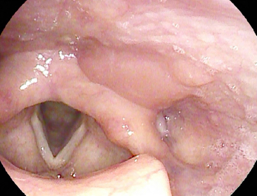
Fig.┬Ā2
Computed tomography (CT) image of the mass in the hypopharynx. CT image shows the mild bulging contour at the left posterior wall of hypopharynx. The margin of lesion is a little enhanced, but there was no peripheral infiltration or abnormally enlarged lymph nodes.
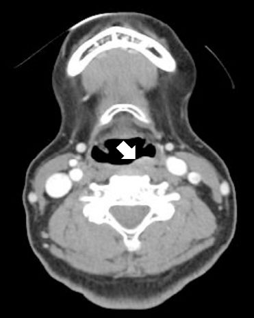
Fig.┬Ā3
Histopathological findings of Mucosa-Associated Lymphoid Tissue lymphoma (MALT lymphoma) in the hypopharynx. (A) Diffuse lymphoid follicular cell proliferation is shown. (Hematoxylin & Eosin, x 100). (B, C) Infiltration of epithelial structures by lymphoid cells (Lymphoepithelial lesion) is shown. Plasma cells with abundant cytoplasm and hyperchromatic nuclei are shown in the marginal zone (Hematoxylin & Eosin, x 400). (D) Immuno-histochemistric finding is shown positive in CD 20 stain.

Ļ│Āņ░░
WHO ļČäļźśņŚÉ ļö░ļź┤ļ®┤ ļ│ĆņŚ░ļČĆ BņäĖĒż ļ”╝Ēöäņóģ(Marginal Zone B cell lymphoma, MZL)ņØĆ ņĀÉļ¦ē ņŚ░Ļ┤Ćņä▒ ļ”╝ĒöäņĪ░ņ¦ü ļ░Å ļ”╝ĒöäņĀł ņÖĖ ļ│ĆņŚ░ļČĆ BņäĖĒż ļ”╝Ēöäņóģ(Extranodal MZL or MALT lymphoma), ļ╣äņן ļ│ĆņŚ░ļČĆ BņäĖĒż ļ”╝Ēöäņóģ(Splenic MZL), ļ”╝ĒöäņĀł ļ│ĆņŚ░ļČĆ BņäĖĒż ļ”╝Ēöäņóģ(Nodal MZL) ļō▒ 3Ļ░Ćņ¦ĆļĪ£ ļČäļźśĒĢĀ ņłś ņ׳ļŗż. ņĀÉļ¦ē ņŚ░Ļ┤Ćņä▒ ļ”╝ĒöäņĪ░ņ¦ü ļ”╝ĒöäņóģņØĆ 1983ļģä IsaacsonĻ│╝ WrightņŚÉ ņØśĒĢ┤ ņ▓śņØī ĻĖ░ņłĀļÉśņŚłņ£╝ļ®░,8) ļ”╝ĒöäņĀł ņÖĖ BņäĖĒż ļ╣äĒśĖņ¦ĆĒé© ļ”╝ĒöäņóģņØś ĒĢśļéśļĪ£, ņŻ╝ļĪ£ ņ£äņŚÉņä£ ļ░£ņāØĒĢśņ¦Ćļ¦ī ņ╣©ņāś, Ļ▓░ļ¦ē, ņĢłņÖĆ, ĒøäļæÉ, ĒÅÉ, ņ£Āļ░®, ņŗĀņן, Ļ░ä ļō▒ ļŗżņ¢æĒĢ£ ņĪ░ņ¦üņŚÉņä£ ļ░£ņāØĒĢĀ ņłś ņ׳ņ£╝ļ®░, ļæÉĻ▓ĮļČĆņŚÉņä£ ļ░£ņāØĒĢśļŖö ļ╣äĒśĖņ¦ĆĒé© ļ”╝ĒöäņóģņØś 7-9%ņŚÉ ĒĢ┤ļŗ╣ĒĢśĻ│Ā ļŗżļźĖ ļ╣äĒśĖņ¦ĆĒé© ļ”╝ĒöäņóģĻ│╝ ļŗ¼ļ”¼ ņŚ¼ņä▒ņŚÉņä£ ņ£Āļ│æļźĀņØ┤ ļŹö ļåÆļŗż.9) 2017 WHOņŚÉ ļö░ļź┤ļ®┤ ņ¦äļŗ©ņØä ņ£äĒĢ┤ņä£ļŖö ņĪ░ņ¦üĻ▓Ćņé¼Ļ░Ć ĒĢäņłśņØ┤ļŗż. ņĀÉļ¦ē ņŚ░Ļ┤Ćņä▒ ļ”╝ĒöäņĪ░ņ¦ü ļ”╝ĒöäņóģņØś ņĀäĒśĢņĀüņØĖ ĒŖ╣ņ¦Ģ ņżæ ĒĢśļéśļĪ£, ņżæņŗ¼ĻĄ¼ ņ£Āņé¼(centrocyte-like) ļ│ĆņŚ░ļČĆ BņäĖĒżļŖö ņżæņåī Ēü¼ĻĖ░ņØś ņĢĮĻ░ä ļČłĻĘ£ņ╣ÖĒĢ£ ĒĢĄņØä Ļ░Ćņ¦ĆĻ│Ā ņ׳ņ£╝ļ®░, ĒĢĄņåīņ▓┤ļŖö ļłłņŚÉ ļØäņ¦Ć ņĢŖĻ│Ā, ņŚ╝ņāēņ¦łņØĆ ņĀüļŗ╣Ē׳ ļČäņé░ļÉśņ¢┤ ņ׳Ļ│Ā, ņäĖĒżņ¦łņØĆ ņśģņØĆ ņāēņØä ļØäĻ│Ā ņ׳ļŗż. ļŗżļźĖ ņóģļźśņØś ļ”╝ĒöäņóģĻ│╝ Ļ░Éļ│äņØä ņ£äĒĢ┤ ļ®┤ņŚŁ ņŚ╝ņāēņØä ĒĢśļŖöļŹ░, ĻĘĖ ņóģļźśļĪ£ļŖö ņŻ╝ļĪ£ CD20, CD10, CD5, CD23, Cyclini D1, ļ®┤ņŚŁĻĖĆļĪ£ļČłļ”░ D (IgD), ļ░Å SOX-1 ļō▒ņØ┤ ņ׳ļŗż.10) ļ│Ė ņ”ØļĪĆņØś Ļ▓ĮņÜ░ ļ╣äņĀäĒśĢņĀüņØĖ ļ”╝ĒöäņäĖĒż ņ╣©ņ£żņØ┤ ĒÖĢņØĖļÉśņŚłņ£╝ļ®░, ļ®┤ņŚŁ ņŚ╝ņāēņāü CD20 ņ¢æņä▒, CD3 ņØīņä▒, Ki-67 3% Ļ▓░Ļ│╝ ļéśņÖĆ ļ│ĆņŚ░ļČĆ BņäĖĒż ļ”╝Ēöäņóģ, ņ”ē ņĀÉļ¦ē ņŚ░Ļ┤Ćņä▒ ļ”╝ĒöäņĪ░ņ¦ü ļ”╝Ēöäņóģņ£╝ļĪ£ ņ¦äļŗ©ļÉśņŚłļŗż.
ĒĢśņØĖļæÉņŚÉņä£ ņ¦äļŗ©ļÉ£ ņĀÉļ¦ē ņŚ░Ļ┤Ćņä▒ ļ”╝ĒöäņĪ░ņ¦ü ļ”╝ĒöäņóģņØś ļé┤ņŗ£Ļ▓ĮņĀü ĒŖ╣ņ¦ĢņØĆ ņĢäņ¦üĻ╣īņ¦Ć ļ│┤Ļ│ĀļÉ£ ļ░ö ņŚåļŗż. ļ│Ė ņ”ØļĪĆņØś Ļ▓ĮņÜ░ Ēæ£ļ®┤ņØ┤ ļČĆļō£ļ¤ĮĻ│Ā, ĒāĆņøÉĒśĢņØ┤ļ®░, ņŻ╝ļ│Ć ņĀÉļ¦ēĻ│╝ ļ╣äņŖĘĒĢ£ ņĪ░ņ¦üņ£╝ļĪ£ ļŹ«ņØĖ ņóģĻ┤┤Ļ░Ć ĒÖĢņØĖļÉśņŚłļŗż. ņĀÉļ¦ē ņŚ░Ļ┤Ćņä▒ ļ”╝ĒöäņĪ░ņ¦ü ļ”╝ĒöäņóģņØ┤ Ļ░Ćņן ĒśĖļ░£ĒĢśļŖö ļČĆņ£äņØĖ ņ£äņŚÉņä£ ļé┤ņŗ£Ļ▓Į ņåīĻ▓¼ņØĆ ļČłĻĘ£ņ╣ÖĒĢ£ ļ¬©ņ¢æņØś Ēæ£ņ×¼ņä▒ ļ»Ėļ×Ć, ĻČżņ¢æ, ĒÖĢļīĆļÉ£ ņŻ╝ļ”ä, ļæÉĻ║╝ņøīņ¦ä ņ£äļ▓ĮĻ╣īņ¦Ć ļŗżņ¢æĒĢśĻ▓ī ļ│┤Ļ│ĀļÉśĻ│Ā ņ׳ļŗż.11)
ņ╣śļŻīļŖö ņ£äļ¦ÉĒŖĖļ”╝Ēöäņóģ(Gastric MALT lymphoma)ņØś Ļ▓ĮņÜ░ Helicobacter pylori ņĀ£ĻĘĀ ņ╣śļŻīļź╝ ņ┤łņ╣śļŻīļĪ£ ņŗ£Ē¢ēĒĢśļéś, ņ£äļ¦ÉĒŖĖļ”╝ĒöäņóģņØ┤ ņĢäļŗī Ļ▓ĮņÜ░ ņĀ£ĻĘĀ ņ╣śļŻīļŖö ĻČīņ£ĀļÉśņ¦Ć ņĢŖļŖöļŗż.10) ņ╣śļŻī ļ░®ļ▓ĢņØĆ ļ│æĻĖ░ņŚÉ ļö░ļØ╝ Ļ▓░ņĀĢļÉśļŖöļŹ░, ļīĆļŗżņłśļź╝ ņ░©ņ¦ĆĒĢśļŖö 1, 2ĻĖ░ļŖö ņĀ£ĒĢ£ļÉ£ ļ│æĻĖ░ļĪ£, ĻĄŁņåī ņ╣śļŻī ļśÉļŖö ņĀäņŗĀ ņ╣śļŻīļź╝ ņŗ£Ē¢ēĒĢ£ļŗż.10,12) ĻĄŁņåī ņ╣śļŻīļŖö ņłśņłĀĻ│╝ ļ░®ņé¼ņäĀ ņ╣śļŻīļĪ£ ļéśļłī ņłś ņ׳ļŖöļŹ░, ņłśņłĀņØĆ ņÖäņĀä ņĀłņĀ£Ļ░Ć Ļ░ĆļŖźĒĢśĻ│Ā, ņłśņłĀ Ēøä ņāØĒÖ£ņŚÉ ņśüĒ¢źņØä ņŻ╝ņ¦Ć ņĢŖņØä ņĀĢļÅäņØś ļ▓öņ£äņØĖ Ļ▓ĮņÜ░ņŚÉļŖö ĒÜ©Ļ│╝ņĀüņ£╝ļĪ£ ļ│┤Ļ│ĀļÉśĻ│Ā ņ׳ņ£╝ļéś, ņłśņłĀņØś ļ▓öņ£äĻ░Ć Ļ┤æļ▓öņ£äĒĢĀ Ļ▓ĮņÜ░ņŚÉļŖö ņłśņłĀņĀü ņĀłņĀ£ņŚÉ ņ׳ņ¢┤ ļ│┤ļŗż ļŹö ņŗĀņżæĒĢ£ Ļ▓░ņĀĢņØä ņÜöĒĢ£ļŗż.10,13) ļ░®ņé¼ņäĀ ņ╣śļŻīļŖö ĒŖ╣Ē׳ ņ£äļ¦ÉĒŖĖļ”╝Ēöäņóģ ņÖĖ ļ░£ņāØĒĢ£ ņĀÉļ¦ē ņŚ░Ļ┤Ćņä▒ ļ”╝ĒöäņĪ░ņ¦ü ļ”╝ĒöäņóģņØś 1ņ░© ņ╣śļŻīļĪ£ ņל ņĢīļĀżņĀĖ ņ׳ņ£╝ļ®░ ļ│┤ĒåĄ 25-30Gyļź╝ 10-15ļ▓łņŚÉ ļéśļłäņ¢┤ ņĪ░ņé¼ĒĢ£ļŗż.14) ņĀäņŗĀĒĢŁņĢöņ╣śļŻīļéś ļ®┤ņŚŁņ╣śļŻīļŖö ņל ņŗ£Ē¢ēļÉśņ¦Ć ņĢŖņ£╝ļ®░, ļ░®ņé¼ņäĀ ņ╣śļŻīņŚÉ ĻĖłĻĖ░ņŚÉ ĒĢ┤ļŗ╣ĒĢśĻ▒░ļéś, ļ░®ņé¼ņäĀ ņ╣śļŻīņŚÉ ņŗżĒī©ĒĢśņśĆņØä ļĢī ļō▒ ĻĄŁĒĢ£ļÉ£ Ļ▓ĮņÜ░ņŚÉ ņŗ£ļÅäĒĢ┤ ļ│╝ ņłś ņ׳ļŗż.10,15) ļ│Ė ņ”ØļĪĆņØś Ļ▓ĮņÜ░ ņłśņłĀņØä ĒåĄĒĢ┤ ņĀÉļ¦ē ņŚ░Ļ┤Ćņä▒ ļ”╝ĒöäņĪ░ņ¦ü ļ”╝Ēöäņóģņ£╝ļĪ£ ņ¦äļŗ©ņØ┤ ļÉśņŚłņ£╝ļ®░, ņłśņłĀ Ēøä ļŗ©ņØ╝ ļČĆņ£äņŚÉ ļ░£ņāØĒĢ£ ņĀÉļ¦ē ņŚ░Ļ┤Ćņä▒ ļ”╝ĒöäņĪ░ņ¦ü ļ”╝ĒöäņóģņØ┤ ĒÖĢņØĖļÉśņ¢┤ ņ╣śļŻīļĪ£ 3,060cGy ņäĀļ¤ēņØś ĻĄŁņåī ļ░®ņé¼ņäĀ ņ╣śļŻīļź╝ ņŗ£Ē¢ēĒĢśĻ│Ā, ņØ┤Ēøä 2ļģä ļäśĻ▓ī ņÖäņĀä Ļ┤ĆĒĢ┤ ņāüĒā£ļź╝ ņ£Āņ¦ĆĒĢśĻ│Ā ņ׳ļŗż.
ĻĄŁļé┤ņŚÉļŖö ņĢäņ¦ü ļ│┤Ļ│ĀļÉśņ¦Ć ņĢŖņØĆ ĒĢśņØĖļæÉņŚÉ ļ░£ņāØĒĢ£ ņĀÉļ¦ē ņŚ░Ļ┤Ćņä▒ ļ”╝ĒöäņĪ░ņ¦ü ļ”╝ĒöäņóģņØś ņ¦äļŗ© ļ░Å ņ╣śļŻīļź╝ Ļ▓ĮĒŚśĒĢśņŚ¼ ņØ┤ļź╝ ļ│┤Ļ│ĀĒĢśļŖö ļ░öņØ┤ļŗż.


